Visual processing improves with locomotion
Polack P-O., Friedman J., and Golshani P. (2013) Cellular mechanisms of brain state-dependent gain modulation in visual cortex. Nature Neuroscience 16(9): 1331–1339.
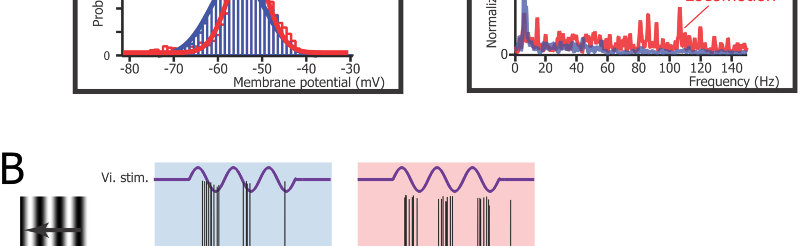
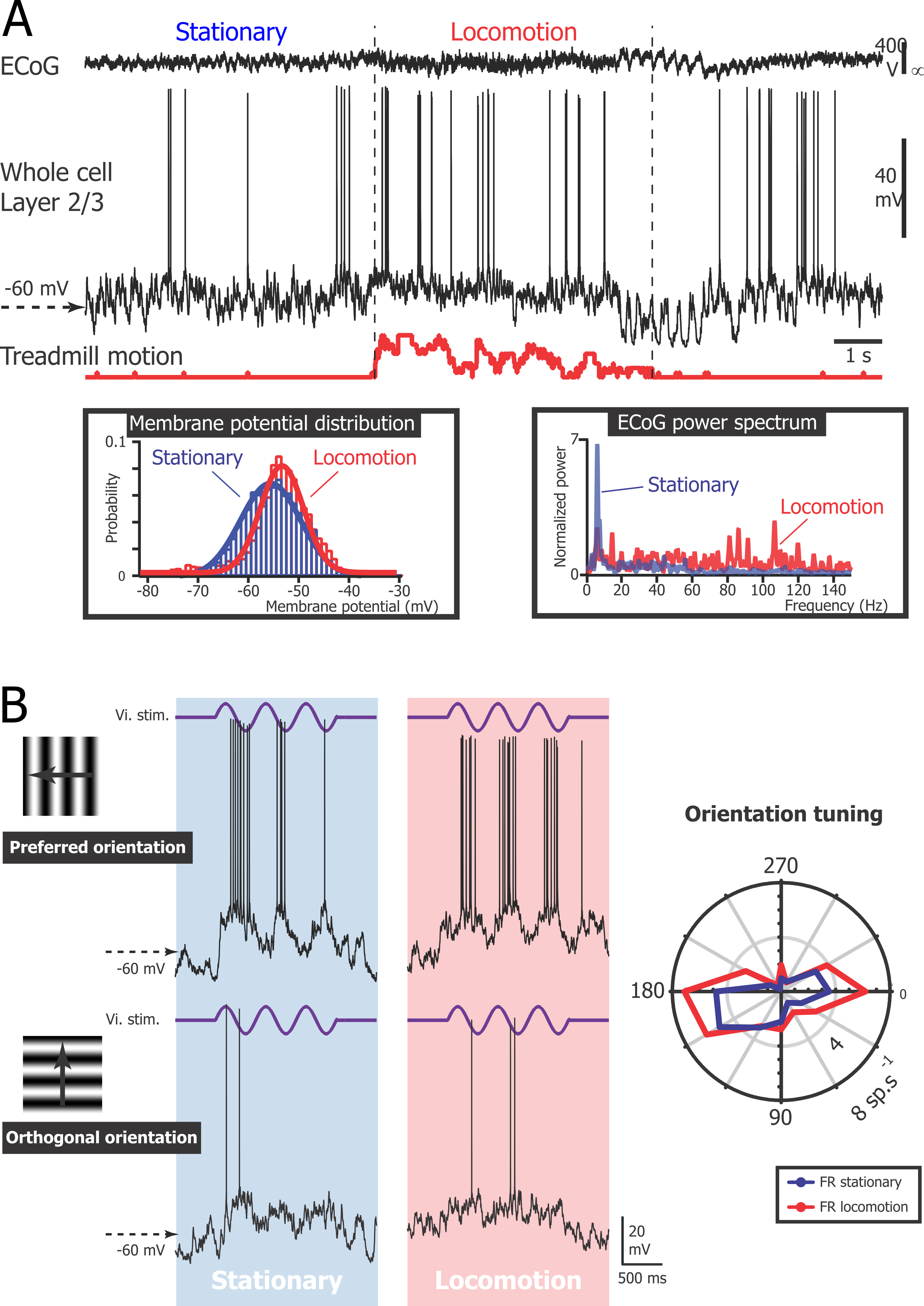
In this article, we show that the neurons located in layer 2/3 and 4 of the primary visual cortex (V1) are more depolarized during locomotion than during the stationary periods. The spontaneous activity (i.e. the action potential discharge when no stimulus is presented) is not affected by this depolarization because the fluctuations of membrane potential decrease in amplitude. However, the response of the neurons to their preferred orientation is enhanced. Our data also suggest that this depolarization is controlled by the noradrenergic system.
Figure
Caption
A. Current-clamp recording of a L2/3 neuron (middle trace) simultaneously with V1 ECoG (top trace) and treadmill motion (bottom trace). Period of locomotion (between vertical dashed lines) was defined as beginning when the velocity was greater than the first step of velocity detection (threshold) for more than 1 s, and resumed when velocity was lower than threshold for more than 1 s. (left inset) Distributions of the membrane potential (Vm ) fitted by Gaussian functions during the periods labeled stationary and locomotion. (right inset) Fast Fourier transforms of the ECoG signal during the periods labeled stationary and locomotion. B. Vm changes (bottom traces) evoked in the same L2/3 neuron by three cycles of a drifting grating at 2 Hz (top traces) of preferred orientation (180 degrees, top panels) and orthogonal orientation (270 degrees, bottom panels) during stationary (left) and locomotion (right) periods. (right inset) Orientation tuning curve of the neuron firing rate (FR) during immobility (blue) and locomotion (red).