An alpha-like rhythm reduces visual processing in V1
Einstein M.C.*, Polack P-O.*, Tran D., and Golshani P. (2017) Visually evoked 3-5 Hz membrane potential oscillations reduce the responsiveness of visual cortex neurons in awake behaving mice. The Journal of Neuroscience. 37(20):5084-5098. * equal contribution.
Low-frequency membrane potential (Vm) oscillations were once thought to only occur in sleeping and anesthetized states. Recently, low-frequency Vm oscillations have been described in inactive awake animals, but it is unclear whether they shape sensory processing in neurons and whether they occur during active awake behavioral states. To answer these questions, we performed two-photon guided whole-cell Vm recordings from primary visual cortex layer 2/3 excitatory and inhibitory neurons in awake mice during passive visual stimulation and performance of visual and auditory discrimination tasks. We recorded stereotyped 3–5 Hz Vm oscillations where the Vm baseline hyperpolarized as the Vm underwent high amplitude rhythmic fluctuations lasting 1–2 s in duration. When 3–5 Hz Vm oscillations coincided with visual cues, excitatory neuron responses to preferred cues were significantly reduced. Despite this disruption to sensory processing, visual cues were critical for evoking 3–5 Hz Vm oscillations when animals performed discrimination tasks and passively viewed drifting grating stimuli. Using pupillometry and animal locomotive speed as indicators of arousal, we found that 3–5 Hz oscillations were not restricted to unaroused states and that they occurred equally in aroused and unaroused states. Therefore, low-frequency Vm oscillations play a role in shaping sensory processing in visual cortical neurons, even during active wakefulness and decision making.
Figure
Caption
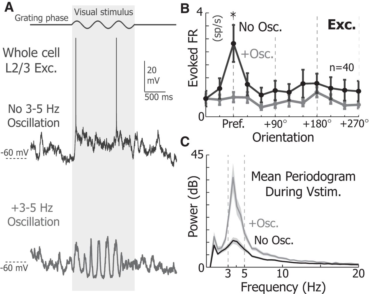
3–5 Hz Vm oscillations reduce the responsiveness of V1 L2/3 neurons. A, Example response of an V1 L2/3 excitatory neuron to a 1.5 s full-screen drifting grating with (bottom) and without (top) a 3–5 Hz oscillation while stimulated at the preferred orientation. B, The mean orientation tuning of excitatory neurons (n = 40) in the presence (gray) and absence (black) of 3–5 Hz Vm oscillations. The firing rate at the preferred angle was significantly larger in the absence of oscillations for excitatory neurons (WRST, p = 8.1e-5). C, The mean periodogram of the Vm during visual stimulation when a 3–5 Hz Vm oscillation is present (gray) and absent (black). Shading represents SEM. *p < 0.05.