Ethosuximide normalize the activity of ictogenic neurons
Polack P-O., and Charpier S. (2009) Ethosuximide converts ictogenic neurons initiating absence seizures into normal neurons in a genetic model. Epilepsia. 50(7): 1816–1820.
The neuronal process by which ethosuximide (ETX), a first choice anti-absence drug, prevents absence seizures is still unresolved. In this article, we made simultaneous in vivo electrocorticographic and intracellular recordings from the cortical focus of the genetic absence epilepsy rat from Strasbourg and examined the effects of systemic injection of ETX at a therapeutic concentration. We show that the interruption of seizures by ETX is correlated with a recovery, in the hyperactive focus neurons, of physiologic values of membrane potential, firing rate, and pattern, as measured in analogous neurons from nonepileptic rats. These data suggest that the anti-absence action of ETX results from the conversion of ictogenic cortical neurons into normal cortical neurons.
Figure
Caption
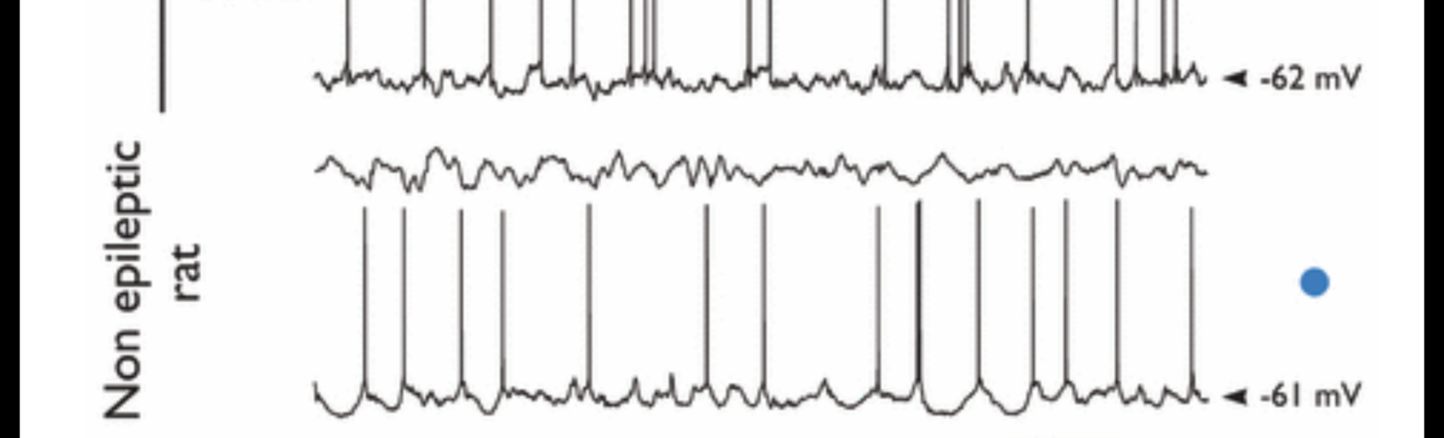
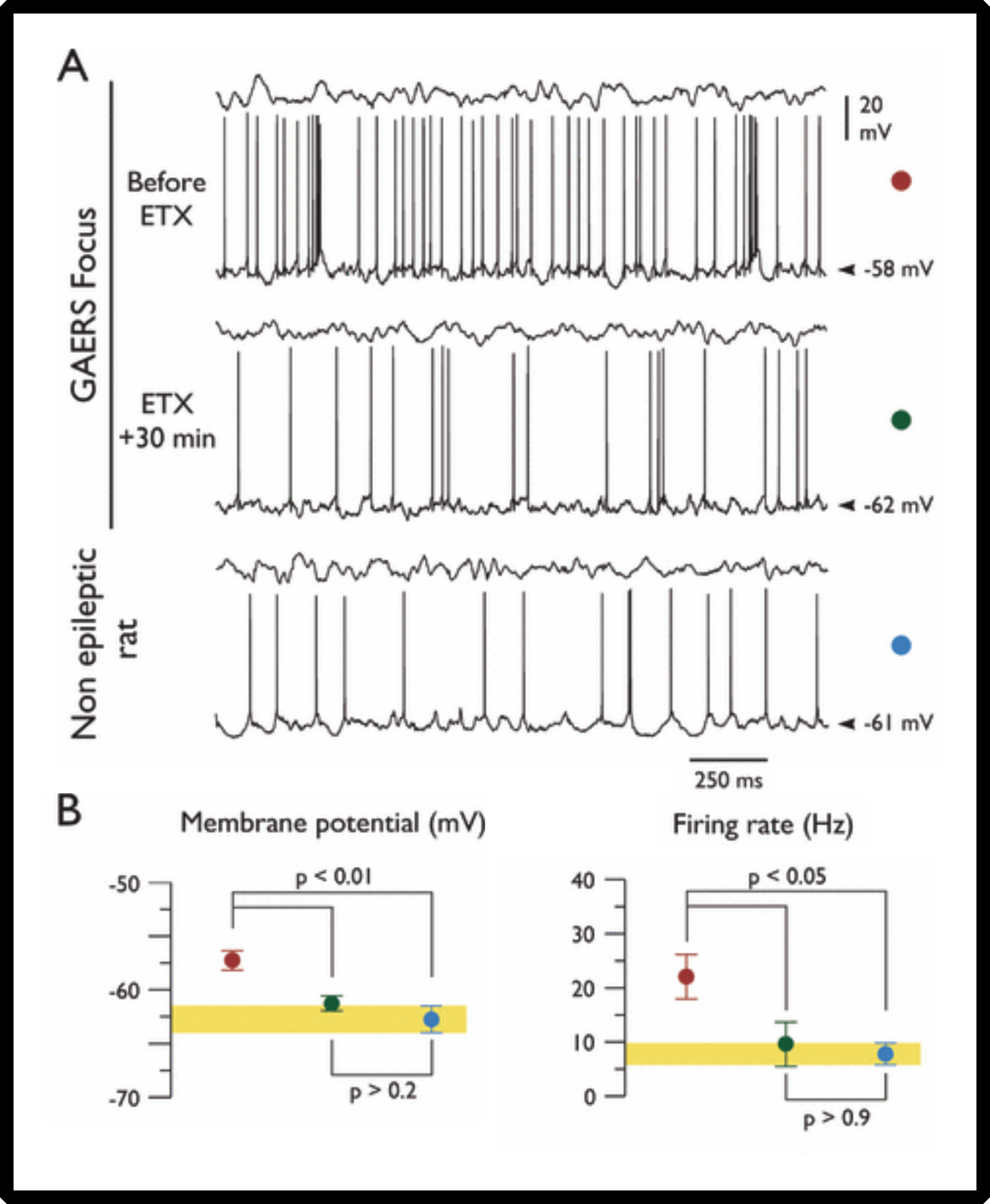
Conversion of hyperactive ictogenic neurons into normal neurons after ethosuximide (ETX) injection. (A) Each paired record shows the electrocorticogram (ECoG) (top trace) and the corresponding interictal intracellular activities (bottom trace) recorded in a layer V neuron from GAERS focus, before and 30 min after ETX injection, and from a layer V cortical neuron located in the facial somatosensory cortex of a nonepileptic Wistar rat. (B) Pooled values of mean membrane potential, and interictal firing rate in deep‐layer neurons from GAERS focus, before (n = 15 neurons; red circles) and after ETX injection (n = 9 neurons; blue circles), and in deep‐layer somatosensory cortical neurons of nonepileptic rats (n = 7 neurons, green circles). The thickness of the yellow strip represents the standard errors of the values measured from normal rats. All the neuronal parameters measured from the GAERS recovered normal values after ETX treatment.